





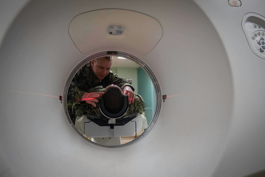
AI in medical imaging isn’t about replacing the experienced human eye, but rather about equipping it with an extraordinary new lens. Essentially, it helps us spot subtle changes and patterns that are incredibly difficult, sometimes even impossible, for a human radiologist or clinician to detect, often due to the sheer volume of data, the minute scale of anomalies, or the complexity of the patterns involved. Think of it as an intelligent assistant that processes vast amounts of visual information at speeds and with an analytical depth that goes beyond human capacity, highlighting areas of concern and offering quantitative insights that dramatically improve diagnostic accuracy and efficiency.
The Foundations of AI in Image Analysis
At its core, AI in medical imaging relies heavily on a branch of artificial intelligence called machine learning, and more specifically, deep learning. This isn’t some futuristic, abstract concept; it’s a sophisticated statistical method that allows computers to learn directly from data without being explicitly programmed for every task.
How Deep Learning Works its Magic
Deep learning uses artificial neural networks, which are inspired by the structure and function of the human brain. These networks are trained on massive datasets of medical images – X-rays, CT scans, MRIs, ultrasound images, and so forth – which have been meticulously labelled by expert radiologists. For example, a network might be fed thousands of lung CT scans, some showing early-stage lung cancer and some healthy, with each scan clearly marked. Through this training, the network learns to identify the characteristic features and patterns associated with the disease. It’s like teaching a child to recognise different animals by showing them countless pictures; eventually, they learn to distinguish a cat from a dog.
The Importance of High-Quality Data
The success of any AI model hinges entirely on the quality and quantity of the data it’s trained on. If the training data is biased, incomplete, or inaccurately labelled, the AI model will inherit those flaws. This is a critical point in medical imaging, as acquiring large, diverse, and expertly annotated datasets is a significant undertaking. Data from various hospitals, different scanning machines, and diverse patient demographics are crucial to ensure the AI can generalise well and perform accurately across a broad patient population, not just the specific group it was initially trained on.
Unpacking What AI Can Detect
So, what exactly can AI “see” that a human might miss? It’s not about magic, but about pattern recognition at a scale and speed that is simply beyond human capability.
Subtle Lesions and Early Disease Detection
One of the most profound benefits is in identifying tiny lesions or very early signs of disease that might be overlooked, especially in busy clinical settings. Consider mammography for breast cancer screening. While radiologists are incredibly skilled, an AI algorithm can analyse thousands of images, picking out minute calcifications or architectural distortions that are characteristic of early tumours, sometimes even before they are visible to the human eye or become palpable. This early detection can be life-saving, leading to earlier intervention and better prognoses. Similarly, in retinal imaging, AI can spot early signs of diabetic retinopathy or glaucoma, often before a human ophthalmologist would, preventing irreversible vision loss.
Quantitative Analysis Beyond Visual Assessment
AI moves beyond just “seeing” things; it can quantify them. For instance, in cardiac MRI, AI can precisely measure ventricular volumes, ejection fractions, and myocardial strain, providing objective metrics that are far more consistent and accurate than manual measurements. In oncology, AI can track changes in tumour size and volume over time with remarkable precision, helping oncologists assess treatment response more effectively. This quantitative aspect is vital for monitoring disease progression, evaluating treatment efficacy, and even predicting future outcomes. It provides objective data points that complement the radiologist’s qualitative assessment.
Identifying Complex Patterns and Biomarkers
Some diseases manifest with highly complex or diffuse patterns that are challenging for the human eye to assimilate and interpret across an entire scan. For example, in neuroscience, AI is being used to analyse MRI scans of the brain to identify subtle changes in white matter integrity or grey matter volume that could be indicative of neurodegenerative diseases like Alzheimer’s or Parkinson’s, often years before clinical symptoms become overt. These patterns might involve correlations across hundreds of slices or subtle texture changes that are individually imperceptible but collectively indicative of pathology. AI can also identify imaging biomarkers that might correlate with genetic predispositions or treatment responses, opening doors for truly personalised medicine.
AI’s Role in Enhancing Workflow and Efficiency
Beyond diagnostic accuracy, AI is also proving to be an invaluable tool in streamlining the incredibly demanding workflow of radiology departments. This isn’t about cutting corners, but about optimising resources and ensuring patients get timely, accurate diagnoses.
Triage and Prioritisation of Urgent Cases
Imagine a busy emergency department with hundreds of imaging studies awaiting review. AI algorithms can be deployed to automatically screen incoming scans for critical findings, such as acute strokes, intracranial haemorrhages, or pulmonary embolisms. If the AI detects a high probability of such an urgent condition, it can flag that study for immediate review by a radiologist, effectively triaging the workload. This ensures that life-threatening conditions are addressed without delay, potentially saving lives and improving patient outcomes, without requiring a human to manually review every single scan for urgency.
Reducing Reporting Burden and Standardisation
AI can also assist with the initial drafting of radiology reports or provide standardised measurements. For instance, after an AI has quantified specific features (e.g., tumour volume, organ size), these measurements can be automatically integrated into a draft report. This not only saves radiologists time but also helps to standardise reporting, reducing inter-observer variability – meaning different radiologists might describe the same finding slightly differently. While the radiologist will always review and finalise the report, this automation reduces the manual, repetitive tasks, allowing them to focus on the nuanced interpretation and patient context.
Quality Control and Anomaly Detection
AI can serve as an extra layer of quality control. It can be trained to detect subtle inconsistencies or potential errors in image acquisition itself, for example, identifying artefacts that might obscure important findings or checking if the correct anatomical regions have been scanned. Furthermore, some AI systems are designed to act as a “second reader,” reviewing studies after a radiologist to catch any potentially missed findings, especially in high-volume screening programmes. This doesn’t imply human error is rampant, but rather acknowledges that even the most experienced professionals can benefit from an intelligent assistant, especially when faced with hundreds of images daily.
Current Applications Across Medical Specialties
AI in medical imaging is no longer confined to research labs; it’s increasingly integrated into clinical practice across various specialties.
Radiology and Oncology
This is perhaps where AI has seen the most rapid adoption. In radiology, AI aids in the detection of lung nodules on CT scans, breast lesions on mammograms, and abnormalities in bone X-rays. For oncology, AI assists in tumour segmentation (outlining tumours), tracking their growth or shrinkage over time, and even predicting treatment response based on imaging features. It’s used in prostate MRI for enhanced lesion detection and characterisation, aiding in biopsy guidance. The ability to quickly and accurately quantify tumour burden and response is transforming cancer care.
Cardiology and Neurology
In cardiology, AI is revolutionising the analysis of cardiac MRI and CT scans. It automates the measurement of cardiac chambers, blood flow, and myocardial function, providing precise data crucial for diagnosing heart failure, coronary artery disease, and structural heart defects. For neurology, AI is vital in detecting early signs of stroke, identifying brain haemorrhages, and analysing complex patterns in MRI scans for neurodegenerative diseases. It can segment brain tumours, track their progression, and even help in planning neurosurgery. The speed of AI in stroke imaging, for example, is critical as ‘time is brain’.
Ophthalmology and Pathology
AI has made significant inroads in ophthalmology, particularly in screening for diabetic retinopathy, glaucoma, and age-related macular degeneration from retinal images. These conditions, if caught early, can be managed to prevent blindness. In pathology, while not strictly “imaging” in the traditional sense, AI analyses digitised microscope slides (digital pathology) to detect cancerous cells, grade tumours, and identify microscopic patterns that are predictive of disease aggressiveness or response to therapy. This is particularly impactful as pathologists often face incredibly detailed and complex visual tasks.
The Future Landscape and Ethical Considerations
While the potential of AI in medical imaging is immense, it’s crucial to acknowledge the ongoing developments and the ethical landscape that shapes its implementation.
Towards Personalised Medicine and Predictive Analytics
The future will likely see AI moving beyond just detection to increasingly sophisticated predictive analytics. Imagine an AI system that not only identifies a tumour but also predicts its likely aggressiveness, its response to specific therapies, and even the patient’s long-term prognosis, all based on imaging characteristics combined with other clinical data. This moves us towards true personalised medicine, tailoring treatments to individual patients based on their unique biological profile. AI will also play a role in integrating imaging data with genomics, proteomics, and other ‘omics’ data to create a holistic view of a patient’s health.
The Role of Radiologists in an AI-Enhanced World
It’s a common misconception that AI will replace radiologists. Instead, the consensus is that AI will augment their capabilities, transforming their role. Radiologists will evolve from primary image readers to expert diagnosticians who leverage AI tools, validate their findings, and integrate them into a broader clinical context. Their expertise in rare diseases, complex cases, patient communication, and interdisciplinary collaboration will become even more valuable. AI will handle the high-volume, repetitive tasks, freeing up radiologists to focus on critical decision-making and patient care. It will allow them to be more efficient, accurate, and ultimately, more impactful.
Ethical Concerns and Algorithmic Bias
As with any powerful technology, AI in medicine comes with important ethical considerations. One significant concern is algorithmic bias. If AI models are primarily trained on data from specific demographic groups, they might perform less accurately on patients from underrepresented populations. This could lead to disparities in care. Ensuring diverse and representative training datasets is paramount.
Another ethical consideration is accountability. If an AI system makes an error, who is responsible? The developer? The clinician who used it? Clear regulatory frameworks and guidelines are essential to address these issues. Furthermore, patient data privacy and security are paramount, especially given the sensitive nature of medical information. Transparency in how AI algorithms work, known as ‘explainable AI’, is also crucial. Clinicians need to understand why an AI made a particular recommendation to trust and integrate it effectively into their practice. The ongoing development of robust regulatory frameworks, such as those being established by the MHRA in the UK and the FDA in the US, is critical to ensuring the safe and ethical deployment of these transformative technologies.